


In the neurology department of Reyap Hospital, diagnosis and treatment of adult and pediatric brain-nerve muscle diseases are carried out using clinical experience and the latest technological developments.
Diseases and conditions diagnosed and treated in our clinic
Neurologists manage and treat neurological conditions or problems of the nervous system. Patients usually visit a neurologist in the presence of the following symptoms; coordination problems, muscle weakness, emotional changes, confusion, confusion, dizziness, and sensory problems such as touch, vision, or smell.
Some of the most common diseases and conditions treated in the neurology clinic;
Epilepsy
Stroke
Neuropsychology
Headache
Neuro-oncology
Alzheimer’s
Sleeping disorders
Dizziness and balance disorders
Multiple Sclerosis
Parkinson’s and movement disorders
Nerve and muscle diseases
Cerebrovascular diseases (stroke) can be prevented, especially if they are treated within minutes and hours after they occur. In our 24-hour stroke center, emergency treatment is performed and patients are followed up with neurological intensive care and rehabilitation follow-ups. Also, diagnosis and treatment for secondary prevention are made after stroke.
Diagnosis and treatment of epilepsy are done by preoperative evaluation, long-term video-EEG examination, neuropsychological evaluation, MR, PET-CT, and SPECT tests.
Intraoperative imaging methods, monitoring, and prevention of possible diseases are also used.
Neuropsychology Laboratory: EEG, EMG, Transcranial Magnetic Stimulation (TMS)
Balance Laboratory: Posturography
Neurocognitive Laboratory
Sleep Disorders Laboratory
Neurological procedures and tests
During your first appointment to the neurology department, your medical history is taken, and a physical and neurological examination is performed. The neurological examination tests muscle strength, reflexes, and coordination. Because different disorders can have similar symptoms, your neurologist may need more tests to diagnose. Neurologists can recommend a variety of procedures to help diagnose or treat a condition.
Computed Tomography (CT)
CT scan combines a series of X-ray images taken from different angles around your body and create crop-sectional images of bones, blood vessels, and soft tissue of your body. More detailed results are obtained from a CT scan than X-rays.
CT has many applications, but it is particularly suitable for examining people quickly after trauma such as traumas after car accidents. CT scanning is important for the diagnosis of neurological disease as well as for planning surgery or radiation therapy.
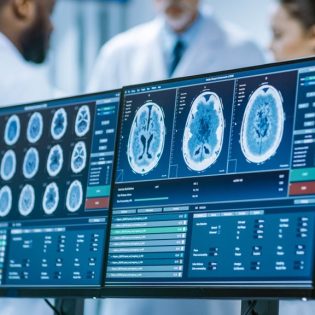
Magnetic Resonance Imaging (MRI)
MRI is a non-invasive way for your doctor to examine your organs, tissues, and skeletal system. It allows obtaining high-resolution images of the inside of the body to help diagnose various problems.
MRI is the most commonly used imaging test in the diagnosis of brain and spinal cord diseases. The most common diseases and conditions diagnosed with MRI are;
Aneurysms of cerebral vessels
Eye and inner ear disorders
Multiple Sclerosis
Spinal diseases
Stroke
Tumors
Trauma-related brain damage
A special type of MRI is functional MRI (fMRI) of the brain. It produces images of blood flow to specific areas of the brain. It can be used to study the anatomy of the brain and determine which parts of the brain perform critical functions. This helps identify important areas of language and movement control in the brains of people considered for brain surgery. Functional MRI can also be used to assess the damage caused by a head injury or disorders such as Alzheimer’s disease.
Positron Emission Tomography (PET) Scan
PET scan is an imaging test that helps reveal how your tissues and organs are working. PET scanning involves the use of a radioactive drug to show this activity. This scan sometimes allows diagnosis before symptoms of the disease appear.
The tracer can be injected, swallowed, or inhaled, depending on the organ or tissue being examined. The tracker gathers in areas of your body with a higher chemical activity that usually corresponds to the disease areas. These areas appear as bright spots on a PET scan.
PET scanning is useful in detecting or evaluating a variety of conditions, including many cancers, heart diseases, and neurological disorders. Usually, it is combined with CT and MRI to increase the diagnostic value and create a treatment program.
Transcranial Magnetic Stimulation (TMS)
TMS is a technique of applying magnetic pulses to the brain through a coil. An electric current is transmitted to the coil placed in the head, which causes an electric current in the brain. Different types of coils are used to reveal different magnetic field patterns and the dosage is increased to stimulate deeper areas of the brain.
TMS is used as a diagnostic tool by using it to map behavioral circuits with spatial and temporal precision, and as a therapeutic tool as it can lead to permanent changes in brain function.
Diseases and conditions that can be treated with TMS;
Parkinson’s disease
Depression
Migraine
Pain management
Ringing in the ears (tinnitus)
Besides, stroke rehabilitation can be done.
Your doctor decides on the placement of the coil and the dose to be applied according to your needs. You need to stay awake and alert during the 40-minute session. You may feel some discomfort in the scalp for a short time during and after treatment.TMS is an outpatient method that is non-invasive and does not require anesthesia. There is no recovery period after treatment and you can go directly to your home.
Electromyography (EMG)
EMG measures the electrical activity between the brain or spinal cord and the peripheral nervous system. This nerve is located in your arms and legs and is responsible for muscle tone during movement and rest times. EMGs can help your neurologist diagnose spinal cord disease as well as general muscle or nerve dysfunction.
During this test, the neurologist places small electrodes in your muscles to help measure activity during periods of movement and rest. During EMG recording, nerve conduction velocity (NCV) recording is usually taken. While EMG measures muscle activity, NCV evaluates your nerves’ ability to send the necessary signals that control these muscles.
In total, the average EMG/NCV combination test can take approximately 1 hour or longer. The patients should avoid stimulants such as caffeine and nicotine for a few hours before the test because of the possibility of affecting the results.
Electroencephalogram (EEG)
With electrodes placed on your scalp, EEG measures electrical activity in the brain. It is used to help diagnose brain conditions such as infections, tumors, and injuries, as well as seizures and psychiatric disorders.
Unlike EMG, EEG usually doesn’t cause any discomfort. Since electrodes in the scalp measure small changes in the brain, doctors make some changes in the environment to measure brain signals such as different lighting or sounds.
You should avoid stimulants on test day, just like EMG. The EEG procedure takes about an hour and sometimes the test is done while you are asleep.
Posturography
Posturography is a test battery used to measure postural control in static or dynamic conditions. It is used to measure central nervous system adaptive mechanisms that are involved in balance control during both standing still and different movements.
Due to the complex interactions in the brain regarding posture and balance, many situations are created in posturography, including the eyes open or closed, and the mobile or immobile platform on which the patient stands, and the source of the problem that causes balance and posture problems is investigated.
Neurocognitive Laboratory
Our neurocognitive lab is a world-class research center designed to study major changes in the brain and behavior during lifelong learning. Research assistants conduct studies using neuroscience, psychology, education, and computer science tools.
Our laboratory has a modern technological infrastructure including EEG, eye/head tracking, virtual reality (VR), and functional near-infrared spectroscopy (fNIRS).
The projects we carry out in our laboratory include:
Developing VR-based learning environments and assessments
Developing diagnostic tools to help schools identify students with autism
Working to improve functional assessments of people with Alzheimer’s disease or dementia
Sleep Disorders Laboratory
Our sleep disorders lab performs comprehensive tests to diagnose sleep disorders. Sleep disorders are analyzed by recording your brain waves, blood oxygen levels, heart rate, breathing, and eye and leg movements during sleeping. It also allows your doctor to adjust your treatment plan if you have previously been diagnosed with a sleep disorder.
In our lab, sleep stages and cycles are monitored to determine if or when your sleep patterns are disturbed and why. The patient may recommend this assessment if the doctor suspects one of these diseases;
Sleep apnea or another sleep-related breathing disorder
Restless leg syndrome
Narcolepsy
REM sleep behavior disorder
Sleepwalking
Unexplained chronic insomnia